Figure 5.
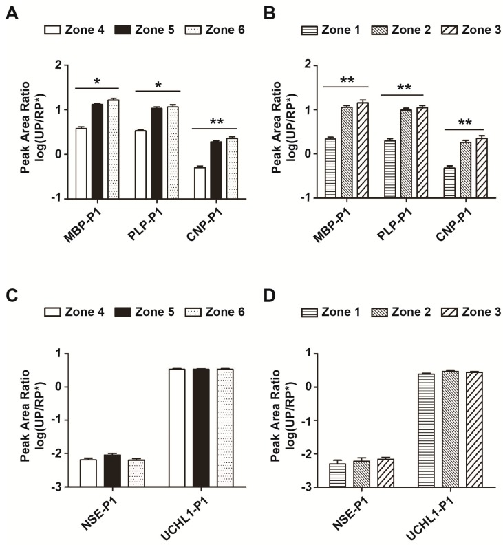
Distribution of myelin proteins and other biomarker candidates in intact and infarcted brain tissue. (A) Three myelin proteins from zones 4–6 of intact brain tissues were verified and semi-quantified by LCM–LRP. Each protein was verified by 2–4 unique peptides and quantified by the peak area. Peak area ratios of unique peptides to RP* were calculated (UP/RP*) (n = 12); (B) Three unique myelin proteins in zones 1–3 of infarcted brain tissue were collected by LCM, and then the peak area ratios of 3 unique peptides to RP* were calculated (UP/RP*) (n = 12); (C) Two protein biomarker candidates from zones 4–6 were verified by 2–3 unique peptides for each and quantified by the peak area. Peak area ratios of unique peptides to RP* were calculated (UP/RP*) (n = 12); (D) The peak area ratios of NSE-P1 and UCHL1-P1 (UP/RP*) in zones 1–3 were calculated (n = 12). Unique peptide 1 for myelin basic protein (MBP-P1), DTGILDSIGR; unique peptide 1 for myelin proteolipid protein (PLP-P1), TSASIGSLCADAR; unique peptide 1 for 2′,3′-cyclic-nucleotide 3′-phosphodiesterase (CNP-P1), AIFTGYYGK; unique peptide 1 for neuron-specific enolase (NSE-P1), LGAEVYHTLK; unique peptide 1 for ubiquitin carboxyl-terminal hydrolase isozyme L1 (UCHL1-P1), FSAVALCK. * p < 0.05; ** p < 0.01.

