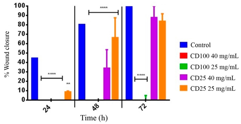
Figure 4.
Wound healing assay displaying percentage wound closure of dermal fibroblast cells in an IBIDI insert cell culturing system at 24, 48, and 72 h. HDFa cells were treated with (red) 40 mg·mL−1 CD-100, (green) 25 mg·mL−1 CD-100, (purple) 40 mg·mL−1 CD-25 and (orange) 25 mg·mL−1 CD-25. Untreated cells served as a control. Data are presented at mean ± SD statistically significant groups were determined by 2-way ANOVA followed by a Bonferroni multiple comparisons test. (** p < 0.01), (**** p < 0.0001) as compared to control.

