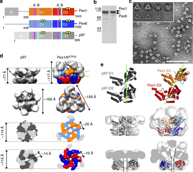
Figure 1. Pex1/6 hexamers are trimers of dimers.
(a) Schematic domain representation of Pex1/Pex6 protomers compared with p97 (N domain, D1/D2 domain). Conserved motifs and residues of each AAA+ domain are indicated: Walker A (A, magenta bars), Walker B (B, turquoise bars), substrate-binding loops (green dots) and arginine finger residues (yellow dots). Non-canonical Walker A and B motifs are indicated as dotted lines. (b) Coomassie-stained SDS–polyacrylamide gel electrophoresis of purified Pex1/6ATP (5 μg, lane 1) or Pex1/6 DWBATP (5 μg, lane 2) overexpressed in E. coli or Saccharomyces cerevisiae. (c) Raw negative stain electron micrograph showing Pex1/6 complexes (40 μg ml−1) incubated with ATPγS. Representative class averages derived from multivariate statistical analysis show top and side views of the Pex1/6ATPγS complex (inset, upper row) and corresponding reprojections of the final 3D reconstruction in the Euler angle directions assigned to the class averages (lower row). Each class contains an average of 5–10 images. Scale bar, 100 nm. (d) Pex1/6ATPγS EM density map as side, top and cross-section views of D1 and D2 rings. Colour code: Pex1 D1 (orange), Pex1 D2 (red), Pex6 D1 (pale blue), Pex6 D2 (blue) and Pex1/6N domains (grey). Equivalent views of p97 (pdb-ID: 3CF3) filtered to 20 Å are shown for comparison. p97 single subunits are coloured alternately light and dark grey. Cross-section viewing planes are indicated by green lines. (e) Cartoon representation of a p97 protomer without N domains and of a Pex1 protomer homology model, seen from the side of the complex. Domain offset between Pex1 D1 and Pex1 D2 is indicated by green dotted lines. Walker A and Walker B motifs are shown as spheres and coloured as in a (upper row). Side view of a p97 dimer fitted as a rigid body into low-pass filtered p97 crystal structure and Pex1/6 heterodimer docked to Pex1/6ATPγS 3D map (middle row). Cut-open side views of the low-pass filtered p97 crystal structure with p97 D2 placed into the EM density map and of Pex1/6ATPγS map with fitted Pex1 D2 and Pex6 D2 homology models. Black dotted lines indicate the central channel (lower row).

