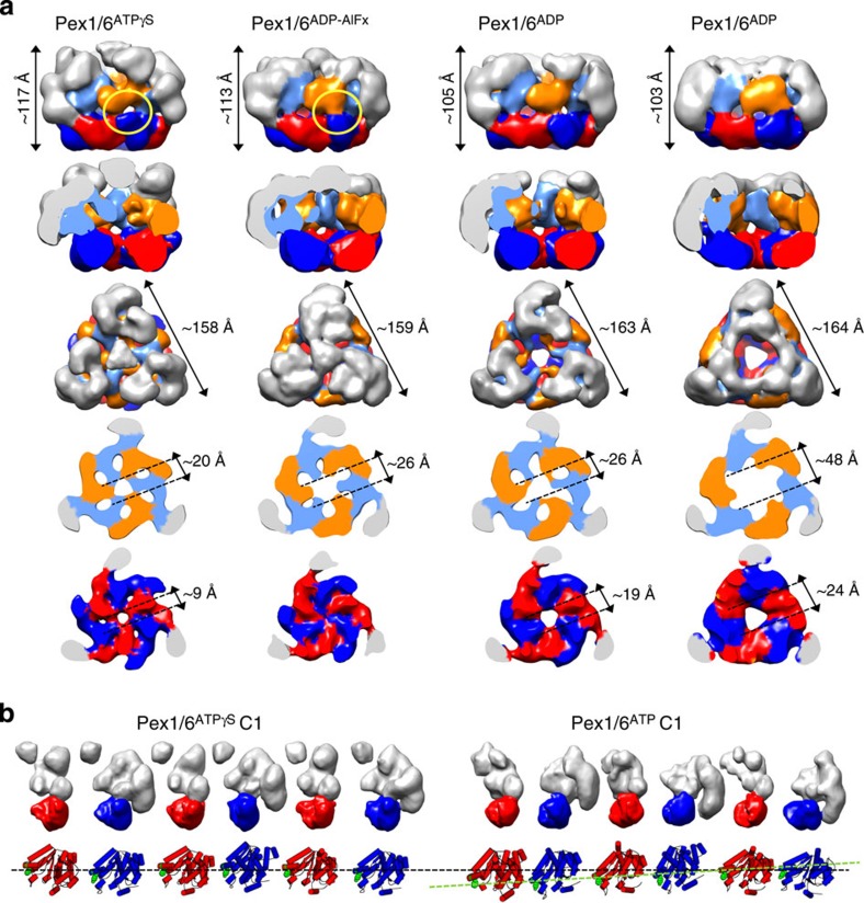
Figure 2. Symmetric and asymmetric wild-type Pex1/6 complexes in the presence of different nucleotides.
(a) EM reconstructions of Pex1/6 complexes in the presence of ATPγS (Pex1/6ATPγS), ADP-AlFx (Pex1/6ADP-AlFx), ATP (Pex1/6ATP) and ADP (Pex1/6ADP) seen as side views (upper row), followed by cut-open side views, top views and cross-sections of the D1 and D2 layers (lower rows). Domain colours correspond to Fig. 1d. (b) Surface representation of each protomer of the asymmetric Pex1/6ATPγS and Pex1/6ATP complex. Pex1 (red) or Pex6 (blue) D2 domains are highlighted. Underneath, a cartoon representation based on rigid body fits of homology models into negative stain EM maps is shown. Pex1F771 and Pex6Y805 are shown as green spheres. The black dotted line indicates the position of pore-facing loops in the D2 domains of asymmetric Pex1/6ATPγS. A green dotted line indicates the asymmetric arrangement of D2 domains in Pex1/6ATP.

