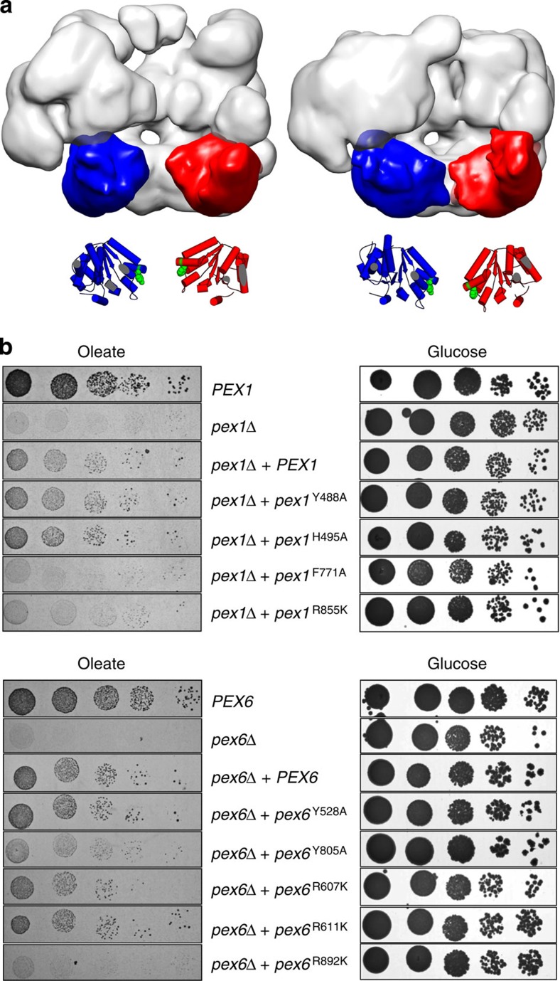
Figure 3. ATP hydrolysis translocates tyrosine loops through movements of D2 domains.
(a) Side-view surface representation of Pex1/6ATPγS and Pex1/6ADP-AlFx. One heterodimer is omitted from the hexamer. One Pex1 D2 and Pex6 D2 domain of each complex is highlighted in red and blue, respectively. Underneath, a cartoon representation based on rigid body fits of homology models into negative stain EM maps is shown. Conserved aromatic residues Pex1F771 and Pex6Y805 are shown as green spheres. (b) Growth of strains expressing either wild-type (PEX1, PEX6), no (pex1Δ, pex6Δ) or mutated pex1, pex6 alleles with a modified pore loop (pex1Y488A, pex1H495A, pex1F771A, pex6Y528A, pex6Y805A) or arginine finger residues (pex1R855K, pex6R607K, pex6R611K, pex6R892K) on either glucose or oleate as a single carbon source.

