
Fig. 1.
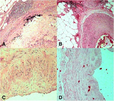
Micrographs showing the coronary tree from patients who died of acute myocardial infarction (AMI) . Panel (a) demonstrates the fatty deposits in adventitia and inflammatory cells surrounding the media of this arteriole are seen (H&E, 200 Ҳ). Panel (b) shows the deposition of lipids and infiltration of lipid-laden foamy cells in the tunica intima and tunica media together with severe narrowing by atherosclerotic plaque, with several hemorrhagic necrotic cores (H&E, 400 Ҳ). Panel (c) indicates histologic finding with fibrous tissue, and necrotic core (H&E, 400 Ҳ). Panel (d) shows microscopic section of the coronary artery shows thickening of the vascular wall by atherosclerotic deposits and hemorrhage. (H&E, 200 Ҳ)