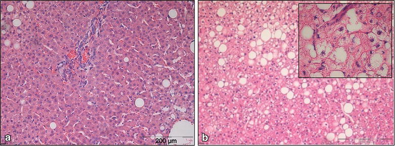
Figure 5.

HE staining of liver samples (magnification 150×). a Liver tissue of a 49 years old patient operated for malignant disease (BMI 28.4 kg/m2) with a low grade fatty degeneration of the liver (<5%) and no lobular inflammation or hepatocyte ballooning. b Liver tissue of a 52 years old patient operated for morbid obesity (BMI 60.0 kg/m2) with grade 2 liver steatosis (40%) and some lobular inflammation and hepatocyte ballooning (top right an enlarged view; magnification 600×).
