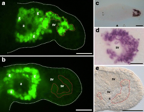
Fig. 7.

RNAi phenotype and ISH pattern of the [0,0,+] class candidate RNA815_80.4. Prostate gland-specific antibody (MPr-1) staining in the tail region of (a) a control worm and (b) a knock-down worm. Note the non-specific antibody staining of the shell/cement glands (g) surrounding the female antrum (a). (c) Whole mount ISH pattern. (d) Detail of the ISH pattern in the prostate glands. (e) Detail of the posterior portion of the tail region of a knock-down worm. White dotted lines show the contour of the animals. Red dotted lines highlight the approximate region of the prostate gland cell bodies. Level of the testes (black triangles) and the ovaries (white triangles); shell/cement glands (g); seminal vesicles (sv); stylet (s). Bars = 100 μm (C), 50 μm (A, B, E), 20 μm (D)
