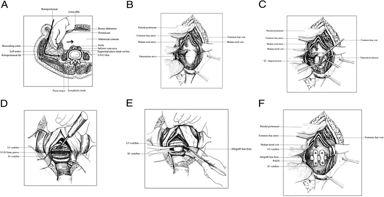
Figure 1.

The surgical procedure used in the study. (A) Exposure of the anterior lumbosacral spine. (B) Ligation of the median sacral artery and vein. (C) An “H”-shaped longitudinal incision and removal of pus. (D) The pus, caseous necrotic tissue, granulation tissue, necrotic bone, and intervertebral disc are removed. (E) Two tricortical allograft iliac bones were trimmed to the proper size and then tightly inserted into the L5–S1 bone groove. (F) Two self-locking titanium anterior lumbosacral vertebrae plates (PACH; General Corp., Germany) of suitable length were selected and anteriorly fixed at L5–S1.
