
Fig. 4.
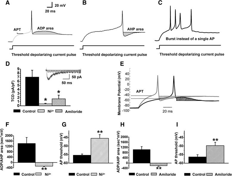
Excitability of the caps−lpH+ neurons is decreased under T-type channel blockers. T-type channel blockers convert the ADP to AHP and significantly increase the AP threshold for the caps−lpH+ neurons at concentrations effectively blocking the Cav3.2 isoform of T-type channels. a,b Action potential parameters used for estimation of changes in excitability of the caps−lpH+ DRG neurons (action potential threshold (APT) and after-depolarization/-hyperpolarization areas (ADP/AHP)). c An example of an AP burst induced by a threshold stimulation in the caps−lpH+ neuron of longer-term diabetic rat instead of a single AP observed in the neurons of normal rats. d Significant decrease of the Ba2+ TCD in the caps−lpH+ neurons under T-type channel blockers Ni2+ (50 μM) and amiloride (1 mM). TCD amplitudes at a voltage step from −100 to −50 mV are presented. Insert: representative traces of Ba2+ current before and after application of 50 μM Ni2+. e A representative example of the AP parameter changes in the caps−lpH+ neurons of diabetic rats under T-type channel blocker, Ni2+ (50 μM). The ADP converted to AHP and the AP threshold increased in 50 μM Ni2+ (grey trace) compared to control (black trace). The ADP and AHP areas are cross-hatched. The AP threshold levels are indicated by dashed lines. f,h The ADP/AHP area reversed from positive to negative values under 50 μM Ni2+ (f) and 1 mM amiloride (h) reflecting conversion of the ADP to AHP shown on (e). g,i Significant AP threshold increase under 50 μM Ni2+ (g) and 1 mM amiloride (i) Numbers of cells: Ni2+ inhibition, n = 6 from three rats; amiloride inhibition, n = 6 from three rats. *p < 0.05, **p < 0.01