
Fig. 6.
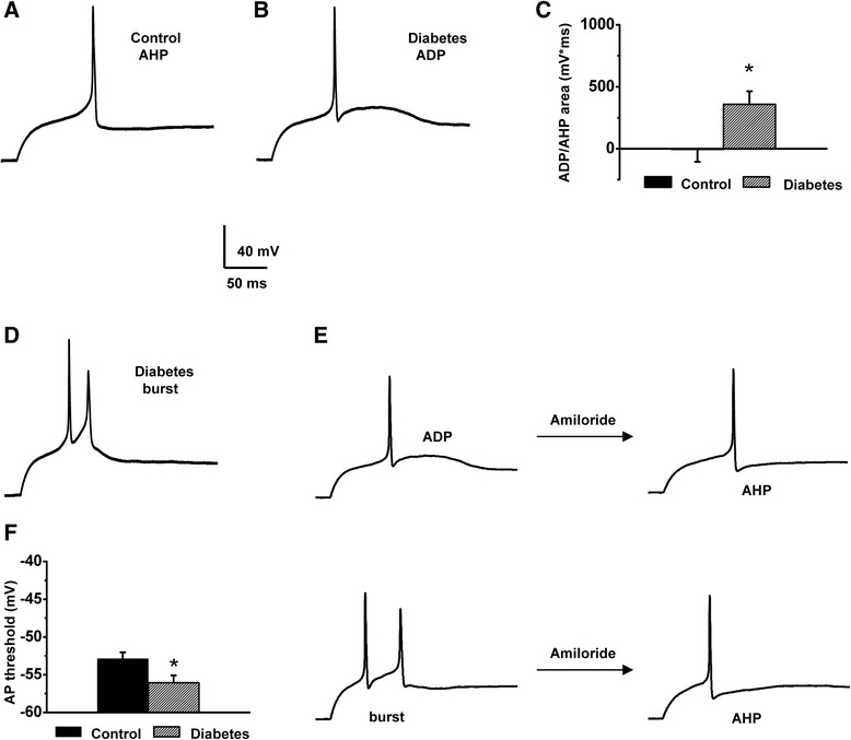
Diabetic-induced changes of the AP parameters for the caps−lpH+ neurons reflect increased excitability of these neurons in diabetes. a A representative AP trace with a slight AHP recorded from the caps−lpH+ neuron of normal rat. b A representative AP trace with a substantial ADP recorded from the caps−lpH+ neuron of diabetic rat. c The ADP/AHP area is significantly increased in diabetes compared to control indicating an increase in excitability of the caps−lpH+ neurons including probability of their bursting in diabetic conditions. Numbers of cells: control for diabetes n = 20 from four rats, diabetes n = 28 from four rats, p < 0.05. d A representative trace of AP bursts generated in 25 % of diabetic neurons (n = 28 from four rats) at a threshold current stimulation. At the same time no AP bursts were observed in the caps−lpH+ neurons of control rats (n = 20 from four rats). e Application of amiloride converts ADPs (top left) and bursts (bottom left) observed in the diabetic caps−lpH+ neurons to AHPs (top and bottom right). f Diabetes leads to a statistically significant decrease in the AP threshold. Numbers of cells: control for diabetes n = 20 from four rats, diabetes n = 28 from four rats, p < 0.05