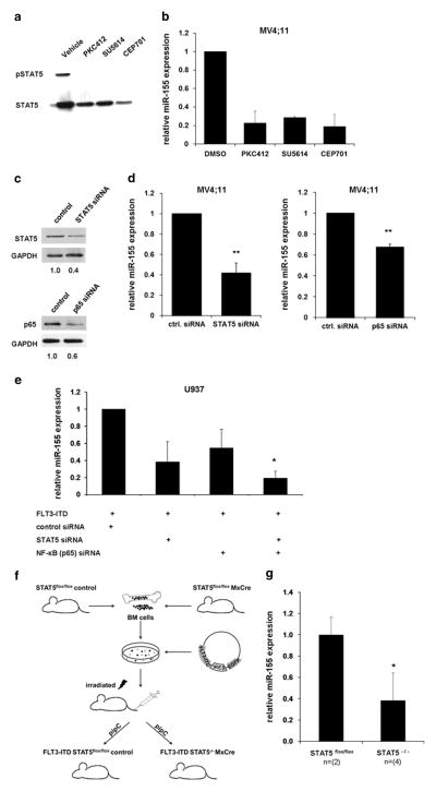
Figure 2.
Block of FLT3-ITD signaling reduces miR-155 expression. MV4;11 cells were treated with PKIs PKC412 (100 nM), SU5614 (1 μM) and CEP701 (100 nM) for 24 h. (a) Western blot shows decreased STAT5 phosphorylation after FLT3-ITD kinase activity inhibition. (b) Expression of miR-155 is decreased after PKI treatment of MV4;11 cells for 24 h. (c) Knockdown of STAT5 or p65 reduces miR-155 expression. Western blot on lysates from MV4;11 cells transfected for 24 h with STAT5 or p65 siRNA. Glyceraldehyde 3-phosphate dehydrogenase (GAPDH) was used as loading control. (d) Expression of miR-155 24 h after transfection of siRNA targeting STAT5 or p65 in MV4;11 cells. (e) Knockdown of STAT5 or p65 inhibits FLT3-ITD-mediated miR-155 induction. U937 cells were co-transfected with FLT3-ITD and siRNAs (control, STAT5, p65 and combination of STAT5 and p65 siRNA). After 24 h, miR-155 expression was analyzed by qPCR. (f) Schematic of workflow to generate an Mx-Cre-inducible STAT5flox/flox knockout mouse model constitutively expressing FLT3-ITD. (g) Analyses of miR-155 expression in bone marrow of FLT3-ITD-inducible Mx-Cre STAT5flox/flox knockout mouse model without and with STAT5 knockout. All values were normalized to U6 (**P ≤ 0.01; *P ≤ 0.05).

