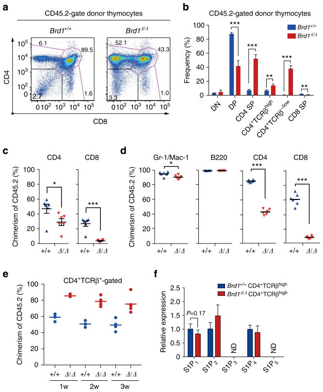
Figure 2. Cell autonomous defect of T cells in the absence of Brd1.
(a) Surface expression of CD4 and CD8 on Brd1Δ/Δ cells in recipient mice. BM cells from Tie2-Cre control (Brd1 +/+) and Tie2-Cre;Brd1fl/fl (Brd1Δ/Δ) mice (CD45.2 +) were transplanted into lethally irradiated wild-type recipient mice (CD45.1+) along with the same number of BM competitor cells (CD45.1 +). Representative flow cytometric profiles of CD4 and CD8 expression in CD45.2+ donor-derived T cells in the thymus at 6 months post-transplantation are depicted. The proportion of each gate is indicated. (b) Frequencies of indicated cell populations in CD45.2 + donor-derived T cells in (a). Data are presented as mean±s.d. (Brd1 +/+, n =5; Brd1Δ/Δ, n =5). (c) Contribution of Brd1Δ/Δ haematopoietic cells to the PB T lymphocytes of recipient mice in (a). Chimerism of donor-derived CD45.2+ Brd1Δ/Δ cells in the PB at 12 weeks post-transplantation is shown as mean±s.d. (Brd1 +/+, n =6; Brd1Δ/Δ, n =6). (d) Contribution of Brd1Δ/Δ haematopoietic cells to the PB in recipient mice. BM cells from Tie2-Cre control (Brd1 +/+) and Tie2-Cre;Brd1fl/fl mice (Brd1Δ/Δ) (CD45.2 +) were transplanted into lethally irradiated wild-type recipient mice (CD45.1+) without any BM competitor cells. Chimerism of donor-derived CD45.2 + Brd1Δ/Δ cells in the PB at 12 weeks post-transplantation is shown as mean±s.d. (Brd1 +/+, n =6; Brd1Δ/Δ, n =6). (e) Competitive repopulating capacity of Brd1Δ/Δ peripheral CD4 T cells. CD4 T cells from the Tie2-Cre control and Tie2-Cre;Brd1fl/fl spleen (1 × 10⁶ each, CD45.2) were transferred into sublethally irradiated NOG mice along with the same number of CD45.1 splenic CD4 T cells (competitor cells). Chimerism of CD45.2+ Brd1Δ/Δ CD4 T cells in the PB CD4+TCRβ+ T-cell pool is plotted as dots and mean values are indicated as bars. (f) Expression of S1P receptors in Brd1Δ/Δ CD4 +TCRβhigh thymocytes. Quantitative RT-PCR analysis of the expression of S1P receptor genes in Brd1 +/+ and Brd1Δ/Δ CD4+TCRβhigh thymocytes was performed. Hprt1 was used to normalize the amount of input RNA. ND =not detected. *P<0.05, **P<0.01 and ***P<0.001 (Student’s t-test).

