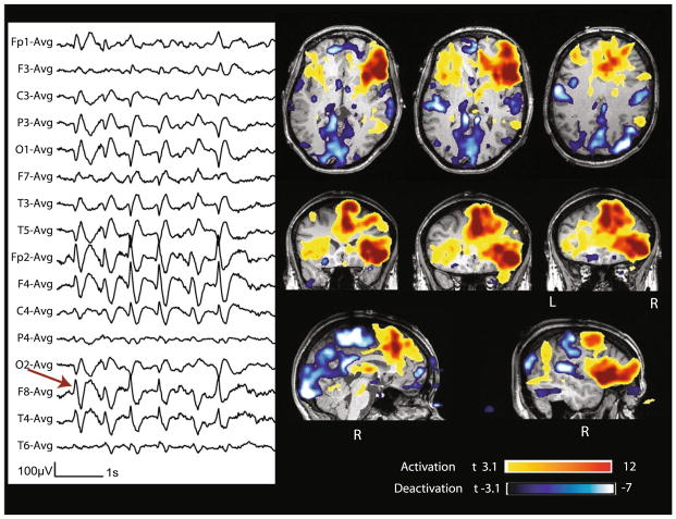
Fig. 5.
Case 22 with lesional right frontal epilepsy (FCD). Marked events: bursts of spike and slow wave complexes with max amplitude at Fp2–F8. BOLD response: max is activation in the right frontal operculum (lesion) and cingulate cortex. Other clusters of activations and deactivations were present, but not investigated because of the lower t-value and because they were remote from the spike field

