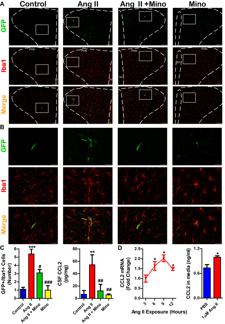
Figure 7. Chronic Ang II infusion increases bone marrow derived microglia/macrophages in the hypothalamic paraventricular nucleus (PVN).
A. Representative images at 10x magnification from the PVN of experimental groups. GFP+ cells are bone marrow derived, and Iba1+ cells indicate microglia/macrophages. Scale bar is 100µm; images taken at Bregma −1.80mm; PVN and third ventricle (3V) are labeled for orientation. B. Higher magnification (40x) images of GFP+/Iba1+ cells in the PVN. Scale bar is 30µm. C. Quantification of GFP+/Iba1+ cells in the PVN reveals an increase in the number of cells in chronic Ang II infusion group, which is decreased by minocycline (mino) treatment. CCL2 content in the CSF was also attenuated by mino (n=5-8 per group). D. Ang II treatment (1µM) of primary hypothalamic neurons induces an increase in CCL2 mRNA and CCL2 protein in the cell culture media. *p < 0.05, **p < 0.01, ***p < 0.001 vs control; #p < 0.05, ###p < 0.001 vs Ang II.

