
Fig. 1. Rapid egress of XNC10-restricted iVα6 T cells from the spleen to the peritoneal cavity following intraperitoneal FV3 inoculation.
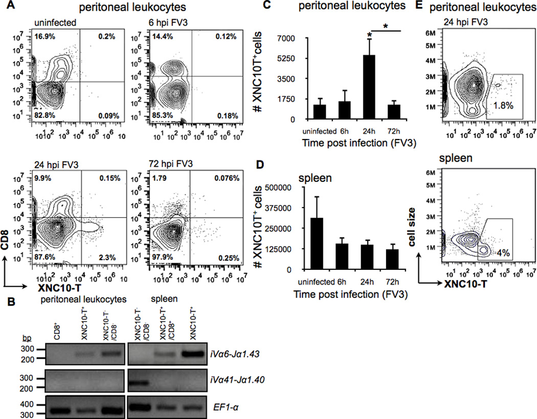
One-year old outbreed X. laevis were infected i.p. with 1 × 10⁶ PFU of FV3, spleen and peritoneal leukocytes were collected at indicated times and analyzed by flow cytometry. (A) Representative flow cytometry of live peritoneal leukocytes isolated by peritoneal lavage from either uninfected or i.p. FV3 infected adults at 6, 24 and 72 hpi and double stained with XNC10-T and anti-CD8 mAb. (B) Rearrangement of specific TCR Vα and Jα genes in the genome of sorted cells from either uninfected spleen of peritoneal leukocytes isolated by peritoneal lavage from FV3 infected adults at 24 hpi. PCR was performed on 50 ng genomic DNA (40 cycles) using primers specific for iVα41-Jα1.40, iVα6-Jα1.43 and Ef1-α Total number of live XNC10-T+ staining cells from either (C) peritoneal leukocytes or (D) spleen leucocytes collected at indicated times. Data are means (± SE) of seven individuals/indicated time point (n=7). *P < 0.05 above bars denotes statistical significance relative to respective uninfected controls and *P < 0.05 above the line denote significant differences between the indicated groups (Student t test). (E) Representative flow cytometry analysis showing forward scatter versus XNC10-T staining for spleen leukocytes and peritoneal leucocytes isolated from either a uninfected frog or 24 hours following FV3 infection respectively.