
Fig. 5. XNC10-deficiency has little effect on the overall recruitment of MHC class I+, MHC class II+ and conventional CD8+ T cells into the peritoneal cavity following FV3 infection.
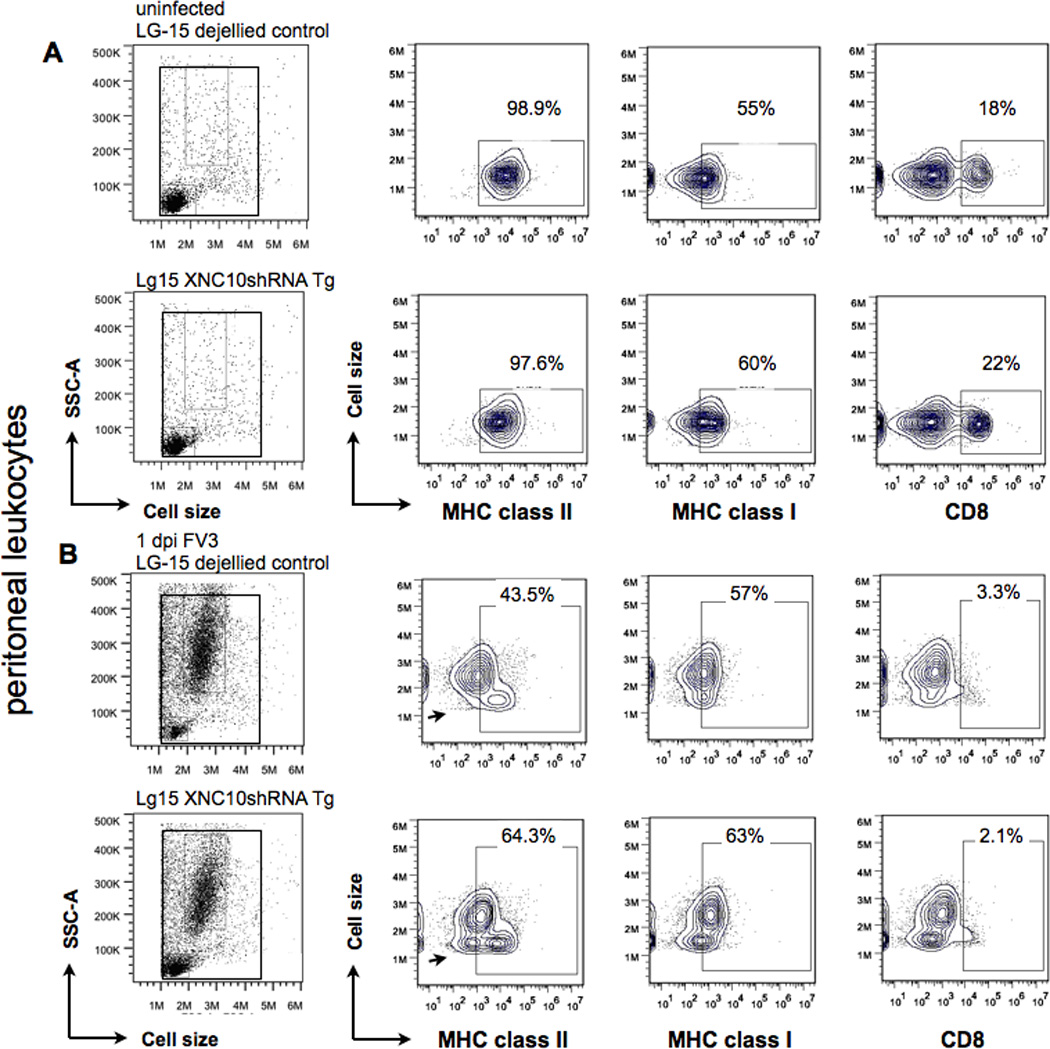
Representative flow cytometry of peritoneal leukocytes isolated from (A) uninfected or (B) FV3 i.p infected (1 × 10⁶ PFU) XNC10-deficient LG-15 transgenic clones and genetically identical non-transgenic LG-15 controls stained with MHC class Ia, MHC class II and CD8-specific mAbs. Scatter profiles and percent positive cells of total live peritoneal are shown. A population of small, agranular MHC class IIlow staining cells found in FV3 infected XNC10-deficient transgenic is indicated by an arrow.