
Fig. 6. XNC10 deficiency results in a delayed antiviral peritoneal leukocyte response.
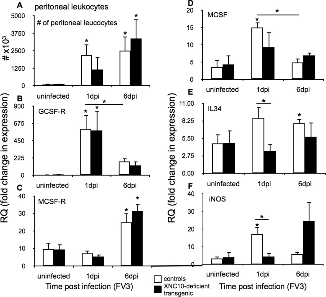
One-year old XNC10-deficient transgenic (n=5, black box) or age-matched dejellied control (n=8, white box) X. laevis were i.p. infected with 1 × 10⁶ PFU FV3. (A) Total number of FV3 induced peritoneal leukocytes. Quantitative gene expression analysis of markers for (B) polymorphonuclear monocytes receptors, GCSF-R, (C) macrophage growth factors receptor MCSF-R (D) M-CSF (E) IL-34 and (F) iNOS. Gene expression was determined relative to a endogenous control (GAPDH) and normalized against respective uninfected control gene expression. All results are presented as mean ± SE and *P < 0.05 denotes statistical significance relative to respective uninfected controls and *P < 0.05, when over bars denotes significant differences between wild-type and XNC10-deficient transgenic groups.