
Fig. 8. Increased viral replication and tissue damages in kidneys of transgenic X. laevis deficient for XNC10 and iVα6 T cells.
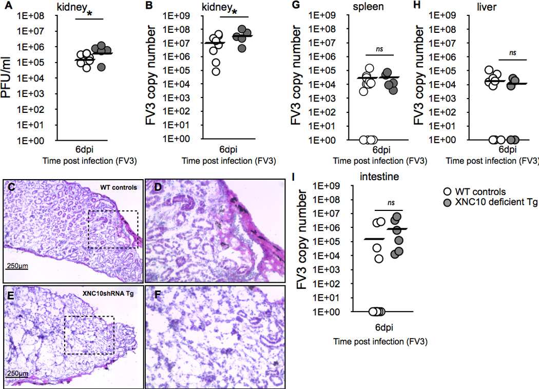
One-year old XNC10-deficient transgenic (grey circles, n=5) or age-matched dejellied control (white circles, n=10) X. laevis where i.p. infected with 1×10⁶ PFU FV3. Kidneys were collected at 6 dpi and divided into three parts (anterior, middle and posterior) to determine (A; middle part) viral loads by absolute qPCR (viral DNA polymerase II, of 625 ng total DNA) and (B; anterior part) plaque assays. *P < 0.05 denotes statistical significance relative to respective uninfected controls and *P < 0.05 denote significant differences between groups denoted by the bars (Student t test). (C–F; posterior part) Representative histology on cryosections stained with hematoxylin and eosin for (C) controls (D) controls at larger magnification, (E) XNC10-deficient transgenic, (F) XNC10-deficient transgenic, larger magnification. (G–I) XNC10-deficiency has little effect on viral dissemination. Viral loads were assessed at 6 dpi in (G) spleen, (H) liver and (I) intestine by qPCR, ns denote no significant differences between groups (Student t test).