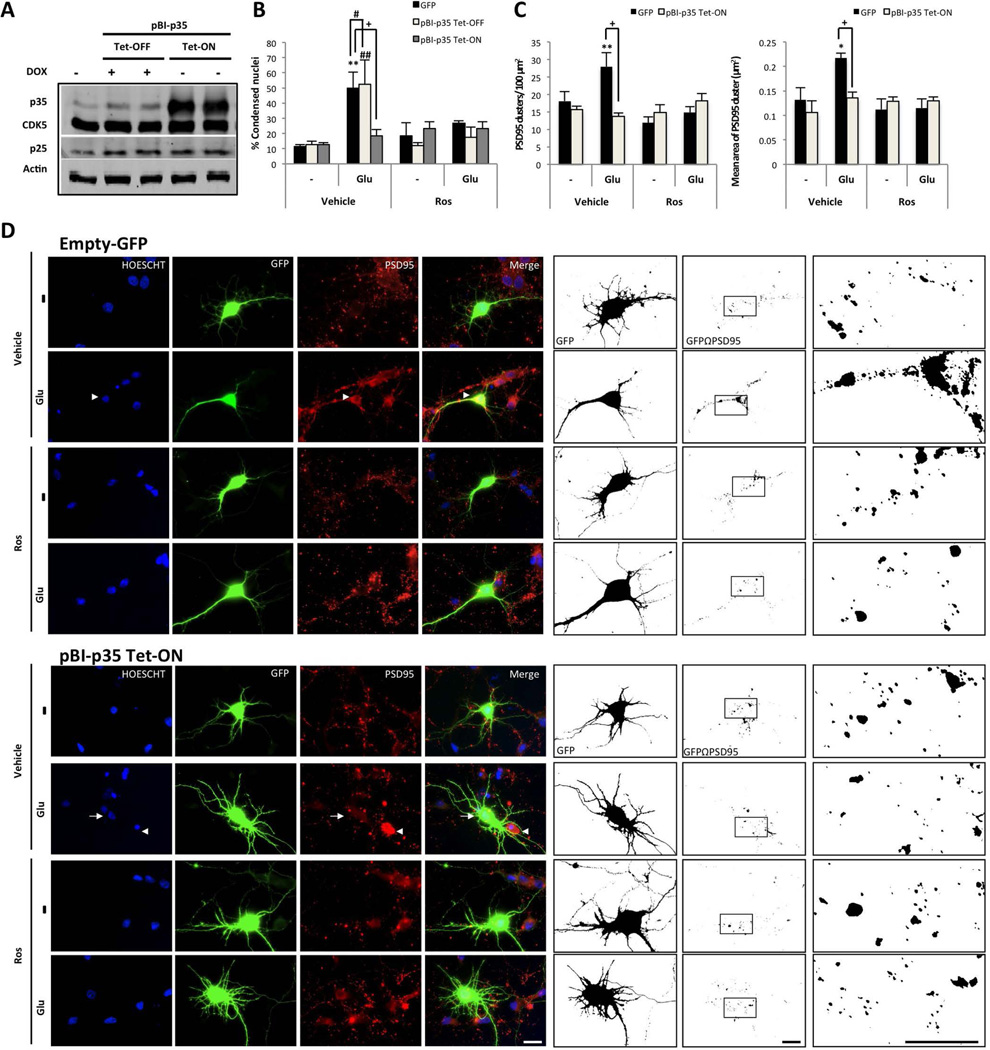
Figure 4.
p35 overexpressed and CDK5 inhibition protects against glutamate excitotoxicity. (A) Hippocampal neurons (DIV5) were co-transfected with pBI-p35/eGFP –tTA, and (B) the percentage of condensed nuclei relative to the total nuclei from the transfected neurons were treated (DIV7) with 125 µM glutamate for 20 min, followed by 10 µM Ros for 24 h. p35 and CDK5 protein levels were detected by Western blotting; β-actin was used as the loading control. eGFP: black bars, Tet-OFF: grey bars and Tet-ON: bright grey bars. (C) The number and area of PSD95 clusters in transfected neurons. The data are presented as the means ± SEM of n = 3, by duplicate. *, #, or + represent the comparisons between each treatment of: eGFP, Tet-OFF and Tet-ON, respectively. *, # or + p<0.05, **, ## or ++ p<0.01, ***, ### or +++ p<0.001; ANOVA with Tukey’s test. (D) The morphological characteristics are shown for the neurons transfected with GFP-tagged plasmids (green), empty-GFP and pBI-p35 Tet-ON. Nuclei were stained with Hoechst (blue), and PSD95 was labelled with Alexa 594 dye (red). The arrowhead shows the condensed nucleus compared with the normal nucleus (arrow). Magnification, 60×. Scale bar, 20 µm. Binary images were used to determine the number and area of the PSD95 clusters on GF- positive neurons. Magnification, 60× with 50% zoom. Scale bar, 20 µm. n=3 per duplicate.

