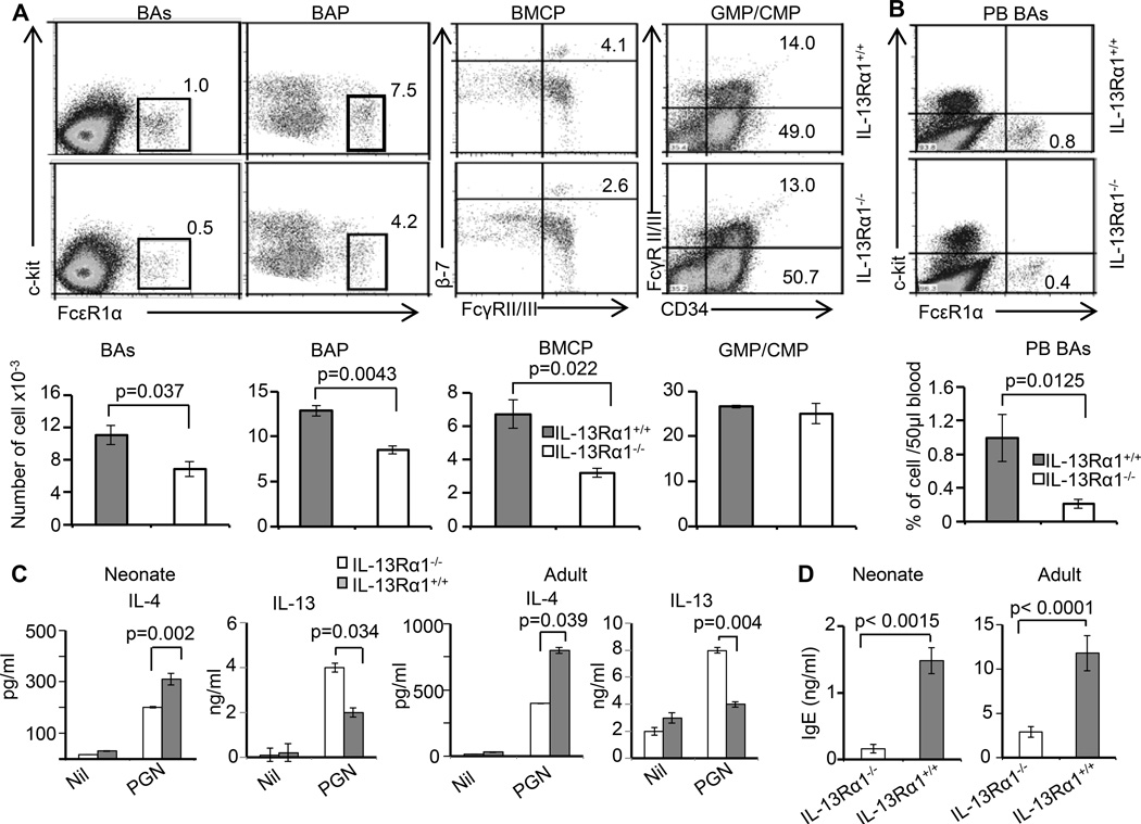
Figure 3.
IL-13Rα1 ablation reduces the frequency of neonatal basophils and interferes with the production of IL-4 cytokine and serum IgE. (A) The top panel shows the frequency of basophils (BAs), basophil progenitor (BAP), basophil/mast cell common progenitors (BMCP), common myeloid progenitors (CMP), and granulocyte monocyte progenitors (GMP) in the bone marrow of IL-13Rα1−/− and IL-13Rα1+/+ neonates. The bar graphs in the bottom panel show the mean ± SD of compiled cell frequencies from 3–5 neonates. (B) The top panel shows the frequency of basophils in peripheral blood (PB BAs) and the bottom panel shows the mean ± SD of compiled cell frequencies from 3–5 neonates. (C) Shows IL-4 and IL-13 production by basophils from neonate (left panel) and adult (right panel) IL-13Rα1−/− and IL-13Rα1+/+ mice upon stimulation with peptidoglycan, a TLR2 ligand. The bars represent the mean ± SD of triplicate wells. The results are representative of 4 experiments with 2–4 mice per group. (D) Shows serum IgE levels in neonatal and adult IL-13Rα1−/− and IL-13Rα1+/+ Balb/c mice as measured by ELISA. Each bar represents the mean ± SD of triplicate wells.

