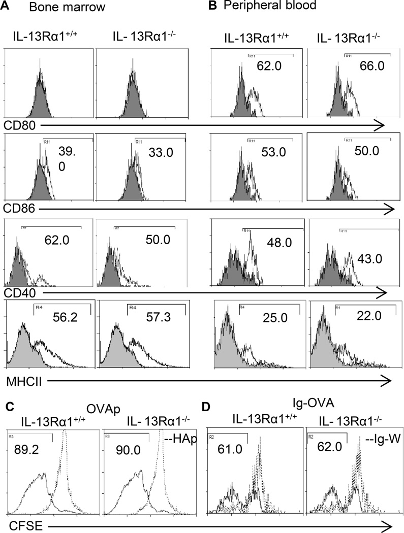
Figure 5. Both IL-13Rα1−/− and IL-13Rα1+/+ basophils function as antigen presenting cells.
BM-derived (A) and peripheral blood (B) basophils were stained with anti-CD80, anti-CD86, anti-CD40 and anti-MHC-II antibodies (open histogram) or isotype control (filled histogram) and marker expression was analyzed by flow cytometry on Lin−c-kit+FcεR1α+ gated basophils. Data is representative of 4 experiments. In (C and D) CFSE-labeled DO11.10 CD4+ T cells (1×10⁶/well) were incubated with bone marrow-derived basophils (1×10⁴/well) from IL-13Rα1+/+ or IL-13Rα1−/− in presence of OVAp (C) or Ig-OVA (D) and antigen presentation was measured by CFSE dilution as described in Materials and Methods. This is representative of 3 independent experiments.

