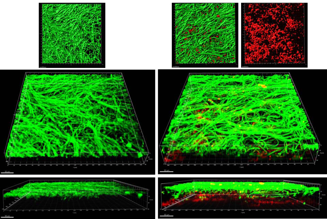
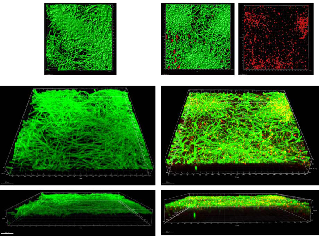
Figure 1.
A: Sixteen-hour mucosal biofilms of C. albicans monospecies (left panel) or C. albicans-streptococci mixed-species (right panel). Biofilms were grown on the surface of three-dimensional models of oral mucosa under wet (media-submerged) conditions. X-Y isosurfaces (top panel) and 3-D reconstructions (bottom panels) of representative confocal laser scanning microscopy images are shown. C. albicans (green) was visualized after staining with a FITC-conjugated anti-Candida antibody. S. oralis (red) was visualized after fluorescence in situ hybridization (FISH) with a Streptococcus-specific probe conjugated to Alexa 546. Scale bar = 50 µm.
B: Sixteen-hour mucosal biofilms of C. albicans monospecies (left panel) or C. albicans-streptococci mixed-species (right panel). Biofilms were grown on the surface of three-dimensional models of oral mucosa under semidry (media limited to inoculum) conditions. X-Y isosurfaces (top panel) and 3-D reconstructions (bottom panels) of representative confocal laser scanning microscopy images are shown. C. albicans (green) was visualized after staining with a FITC-conjugated anti-Candida antibody. S. oralis (red) was visualized after fluorescence in situ hybridization (FISH) with a Streptococcus-specific probe conjugated to Alexa 546. Scale bar = 50 µm.


