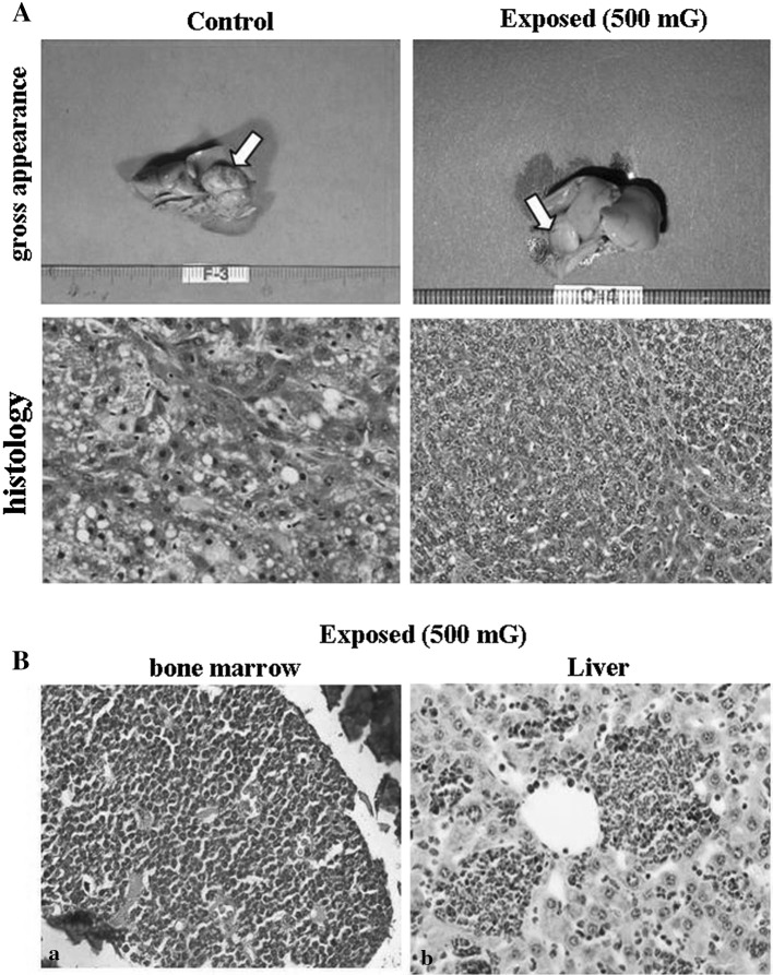
Fig. 3.
Tumors in organ of B6C3F1 mice in the control and exposed group. A Representative images of the liver tumor (gross appearance and histology) in control and EMF exposed mice. Arrows indicate the tumor node. Histology (H&E staining) shows hepatocellular carcinoma (HCC). B Chronic myelogenous leukemia in EMF exposed mice. a Bone marrow, b Liver

