
Fig. 2.
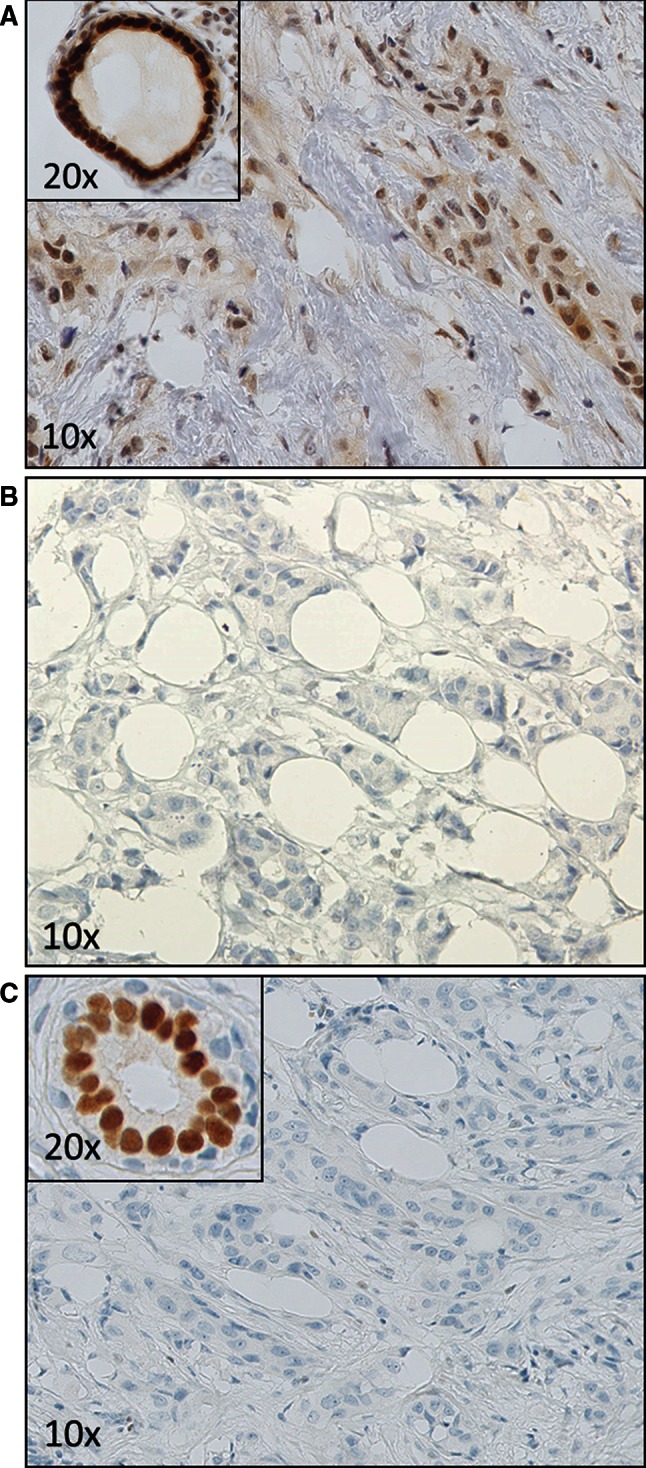
A case where the local result was determined as ER-positive, while this staining was not reproduced on the TMA core and whole-slide testing. A. The local slide which showed both nuclear and smudgy, weaker cytoplasmic staining in the tumor cells as well as associated fibroblasts. A nearby duct is strongly positive. B. The TMA test showing no staining in tumor cells. C. Whole-slide test which verified the ER-negative staining of the TMA, while the normal duct shows an appropriate positive control