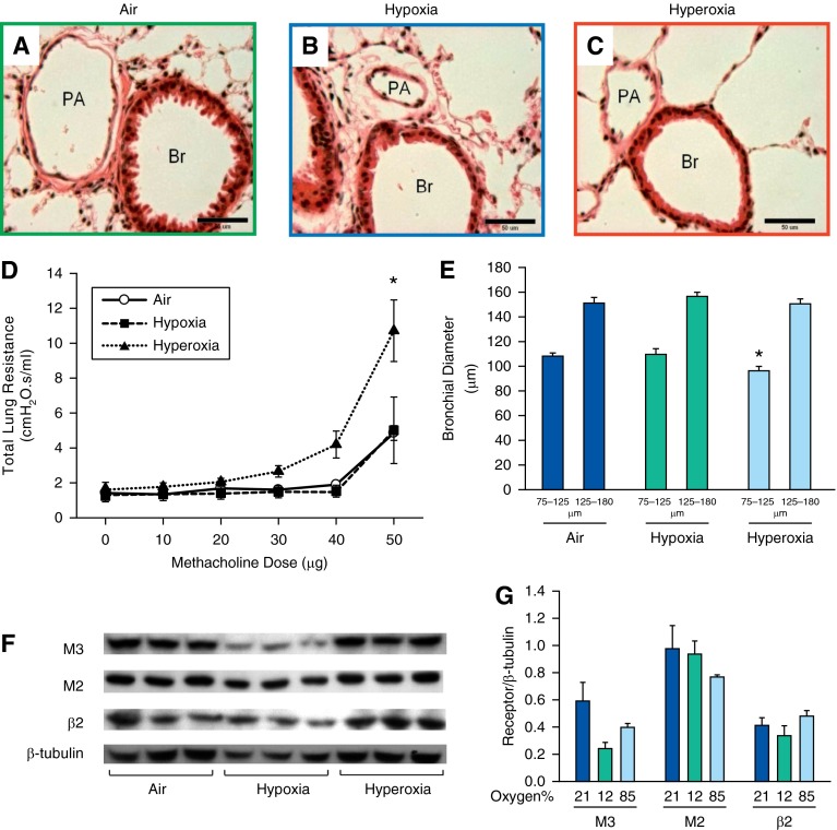
Figure 2.
Airway reactivity was increased in adult mice exposed to neonatal hyperoxia. (A–C) Representative photomicrographs of hematoxylin and eosin (H&E)–stained lung sections of 14-week-old mice exposed to air (A), hypoxia (B), or hyperoxia (C) in the newborn period (400×; calibration bars, 50 μm). Administration of increasing doses of nebulized methacholine (10, 20, 30, 40, and 50 μg) showed an increased airway reactivity only in hyperoxia-exposed mice at the 50-μg dose (D) (n = 21, 7 per group; means ± SE). *P < 0.05 versus corresponding air group. Average diameter of bronchi in the size range of 75–125 μm was decreased in 14-week-old mice exposed to neonatal hyperoxia compared with air control mice (E) (n = 18, 6 per group; means ± SE). *P < 0.05 versus corresponding air group. (F and G) Representative Western blots showing no significant difference in M3, M2, and β2 receptors of lung homogenates of adult mice exposed to air, hypoxia, or hyperoxia in the newborn period (n = 6 per group). Br, bronchiole; PA, pulmonary artery.

