Figure 5.

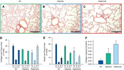
Analysis of lung collagen and elastin content of adult mice exposed to air, hypoxia, or hyperoxia in the newborn period. (A–C) Representative photomicrographs of picrosirius red–stained lung sections of adult mice exposed to air (A), hypoxia (B), or hyperoxia (C) in the newborn period (100×; calibration bars, 250 μm). (D and E) C, collagen-stained area as percentage of total image area; C/T, percentage of collagen area/percentage of tissue area; E, elastin-stained area as percentage of total image area; E/T, percentage of elastin area/percentage of tissue area; TA, tissue area as a percentage of total image area (n = 6 per group; means ± SE). *P < 0.05 versus corresponding air group. Histological quantitation did not show differences in collagen (D) or elastin content (E) among groups. Measurement of soluble collagen in lung homogenates by Sircol soluble collagen assay (F) showed increased collagen in hyperoxia-exposed mice compared with air controls (n = 6 per group). *P < 0.05 versus corresponding air group.
