Figure 2.
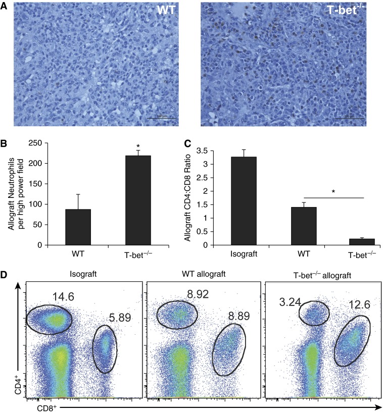
Severe rejection pathology in T-bet−/− recipients is characterized by significantly higher allograft neutrophil count and lower allograft CD4:CD8 ratio compared with WT. (A) Histologic sections of lung allografts from WT and T-bet−/− recipients at Day 10 showing neutrophil staining with antimyeloperoxidase. (B) Total number of neutrophils per high-power field on histological sections of lung allografts stained with antimyeloperoxidase (10 different high-power fields per allograft counted; n = 3 mice per group; P = 0.034; * denotes statistical significance). (C) Lung allograft CD4:CD8 ratios at Day 10 (n = 3–8 mice per group; * denotes statistical significance). (D) Representative flow plots of lung allograft mononuclear cells at Day 10 showing frequencies of CD4+ and CD8+ cells.

