Figure 3.
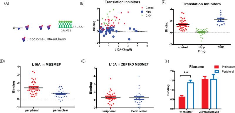
The association of ribosomes with β-actin mRNA in primary MEF. (A) The schematic shows the labeling of large ribosomal subunits by rpL10-mCherry. Primary ZBP1−/− or WT MBS MEF cells were prepared and infected with NLS-tdMCP-EGFP and rpL10A-mCherry (Methods). (B) The association of ribosomes with the mRNA in the presence of translation inhibitors in WT MBS MEF. The number of ribosomes bound per mRNA was plotted as a function of the rpL10A-mCherry concentration. Hippuristanol (Hipp: 1uM/mL) inhibited translation initiation and abolished the association of ribosomes with mRNA. In contrast, cycloheximide (CHX: 2ug/mL) increased the number of ribosomes on mRNA since it slowed down the elongation speed. (C) Summary of the average number rpL10A-mCherry associated with mRNA in the presence of translation inhibitors. Furthermore, FFS experiments were performed in different cellular compartments (D-F). The number of ribosomes binding to β-actin mRNA in the leading edge or perinuclear region was compared in primary MBS MEFs (D) and ZBP1−/− MBS MEF (E). (F) Comparison of ribosome association with mRNA in different cellular regions. There were fewer ribosomes per β-actin mRNA in perinuclear region than in the leading edge in WT MBSMEF. If ZBP1 was knocked out, however, there is no difference in ribosome loading. (*: p<0.05, **: p<0.01, ***: p<0.001). See also Figure S3.

