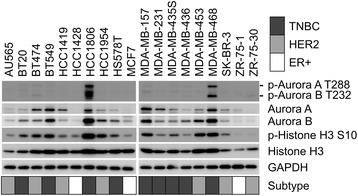
Fig. 4.

Western blot analysis of breast cancer cell lines. Cell lysates from the 19 breast cancer cell lines were subjected to Western blot analysis. Aurora kinases A and B, p-Aurora kinase A and B, and Histone H3 and p-Histone H3 Ser10 were detected using respective antibodies. The experiments were carried out after calibration by using GAPDH as an internal control. Breast cancer subtypes are indicated as follows: gray, TNBC; light gray, HER2; white, ER+
