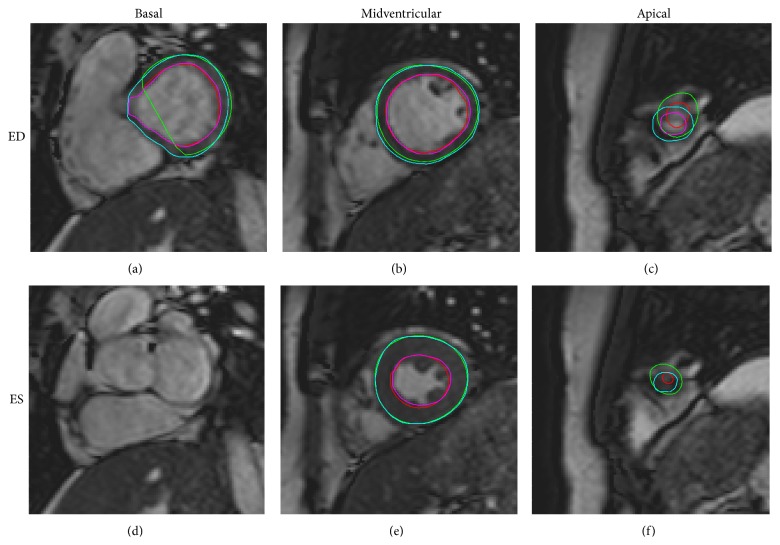
Figure 5.
Automatic segmentation compared to manual delineation in a basal, midventricular and apical slice. Automatic segmentation (endocardium in red and epicardium in green) and manual delineation (endocardium in pink and epicardium in light blue) shown in end-diastole ((a), (b), and (c)) and end-systole ((d) (e), and (f)) for the most basal slice with outflow tract moving out of the imaging plane ((a), (d)), a midventricular slice with papillaries ((b), (e)) and an apical slice with minimal lumen in end-systole ((c), (f)).

