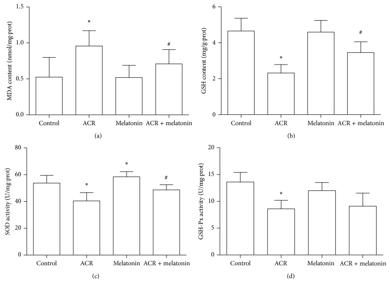
Figure 5.
Effects of MT on MDA generation, GSH depletion, and SOD and GSH-Px activities caused by ACR in rat cerebellum. Experimental animals were divided into four groups: control (equal volume saline) group, ACR (40 mg/kg/day) treatment group, MT (5 mg/kg/day) treatment group, and ACR + MT cotreatment group. Following oral exposure for 12 days, MDA level (a), GSH content (b), SOD (c), and GSH-Px (d) activities were determined by the colorimetric method with a microplate reader. The results are expressed as the mean ± SD (n = 8). ∗ P < 0.05, ∗∗ P < 0.01 versus the vehicle control group, # P < 0.05, ## P < 0.01 versus the ACR treatment group.

