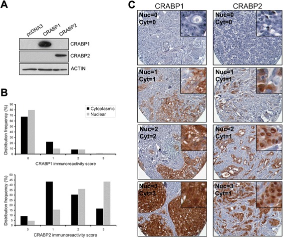
Fig. 2.

Immunoreactivity and subcellular distribution of CRABP1 and CRABP2 in a human primary breast tumor TMA. a Western blot of CRABP1 and CRABP2 in MDA-MB-435 cells transfected with a CRABP1 or CRABP2 expression construct, respectively. b Frequency of breast tumors with different subcellular immunoreactivity scores for CRABP1 and CRABP2: 0, negative; 1, weak; 2, intermediate; 3, strong. c Selected tissue sections from a human breast cancer TMA immunostained with anti-CRABP1 and anti-CRABP2 antibodies. Nuclear (Nuc) and cytoplasmic (Cyt) scores are indicated
