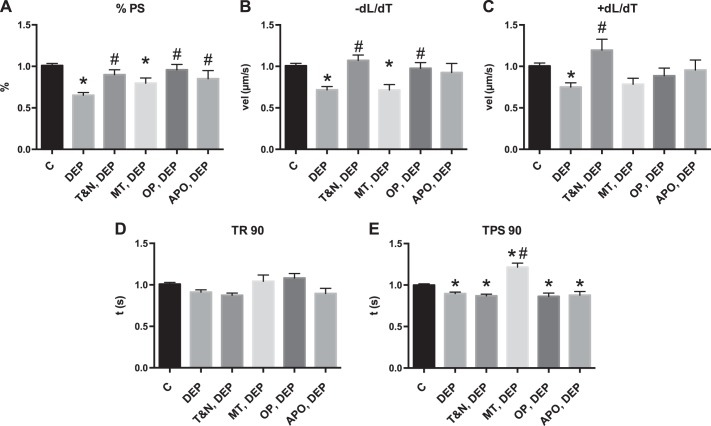
Fig. 2.
Cardiomyocyte contractile dysfunction from DEP treatment is reactive oxygen species mediated. Cardiomyocytes were treated with DEP (25 μg/ml) for 1 h and contractile function measured against control (C). Subsets of cells were pretreated with anti-oxidants N-acetyl-l-cysteine (NAC; 10 mM) and Tiron (10 mM) (T&N), apocynin (APO; 10 mM), mito-tempol (MT; 10 mM), or oxypurinol (OP; 15 mM) for 1 h before DEP treatment A: %PS. B: +dL/dT. C: −dL/dT. D: TR 90. E: TPS 90. All values were analyzed from 8 to 10 cells from 3 to 4 rats each normalized to control cells for each rat. *Significantly different from control treatment; #significantly different from DEP treatment, both P < 0.05 by ANOVA.

