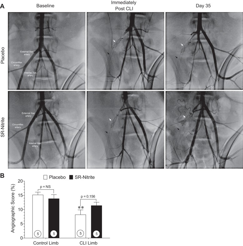
Fig. 6.
Contrast angiographic images and angiographic score at post-CLI day 35. A: magnified-view contrast angiographic images of swine peripheral vasculature at baseline, immediately post-CLI, and at day 35 post-CLI after interventional deployment of a self-expanding endoluminal endoprosthesis consisting of an expanded polytetrafluoroethylene (ePTFE) lining with an external nitinol stent (black arrow) and a self-expanding nitinol mesh occlusion device (white arrow) into the right external iliac artery. B: hindlimb vessel density shown as angiographic score of magnified-view contrast angiographic images of control and CLI limbs distal from the ePTFE-lined endoprosthesis at CLI day 35. Numbers inside circles denote number of animals. **P < 0.01 vs. placebo-treated control limb.

