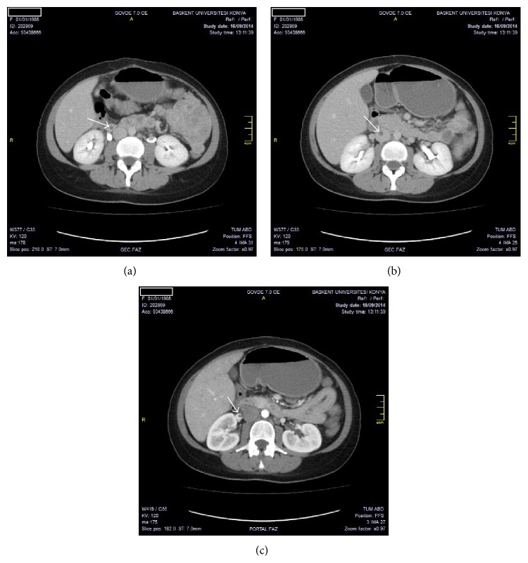
Figure 1.
(a) Ovarian vein thromboses. On axial CT image, hypodense thrombus seen in right ovarian vein. (b) Inferior vena cava vein thromboses. On axial CT image, hypodense thrombus seen in vena cava inferior. (c) Renal vein thrombosis. On axial CT image, hypodense thrombus seen in right renal vein.

