
Figure 9.
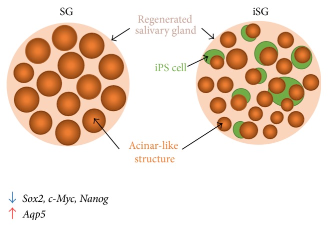
Effect of iPS cells on aggregation of SMG cells. iPS cells (green balls) reduced the size of the epithelial tissue, but acinar-like structure (orange balls) increased their number. iPS cells cannot mix completely with SMG cells and, instead, surround the epithelium of SMG. Stem cell markers, such as Sox2, c-Myc, and Nanog, are decreased, but Aqp5 is increased in iSG.