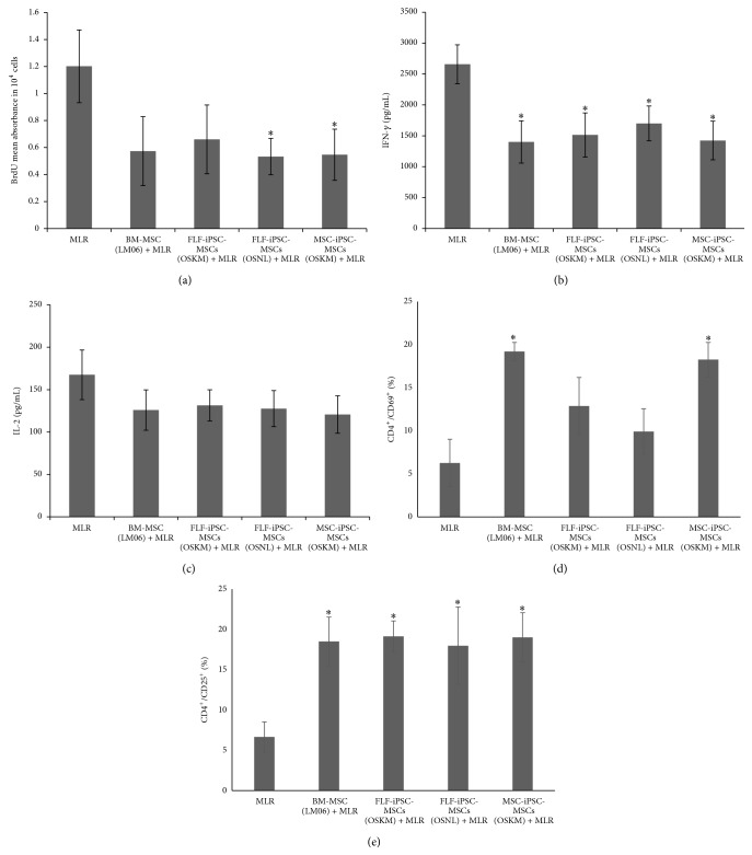
Figure 6.
Status of activated CD4+ T cells in the presence of hiPSC-MSCs. (a) IFN-γ were determined at 48 hours by ELISA. The values are the means ± SD from 3 independent experiments, (b) concentrations of IL-2, and (c) proliferation in MLR/MSC cocultures. MLR cultures were set up in presence or absence of hiPSC-MSCs. BrdU incorporation was significantly lower in MSC-ips-MSCs and FLFiPSC-MSCs (OSNL) in comparison to absence of MSCs. (d) Expression of the T-cell activation markers CD69 and (e) CD25 on CD4+ 5 days after stimulation in a 12-well dish in the presence or absence of hiPSC-MSCs. ∗: significance difference with MLR p ≤ 0.05.

