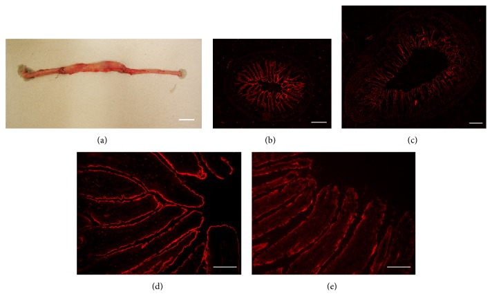
Figure 7.
Macroscopy appearance of interposed ileum (a) and immunohistochemistry of the interposed ileum ((c), (e)) in comparison with the respective ileal segment of a sham-operated animal ((b), (d)). The samples were taken after the animals had been killed in the 3rd week after sham or IIP surgery. Immunostaining in (b)–(e) was performed with SGLT1-Ab. Representative images are shown. Bars: (a) 2 cm, (b) and (c) 500 μm, and (d) and (e) 50 μm.

