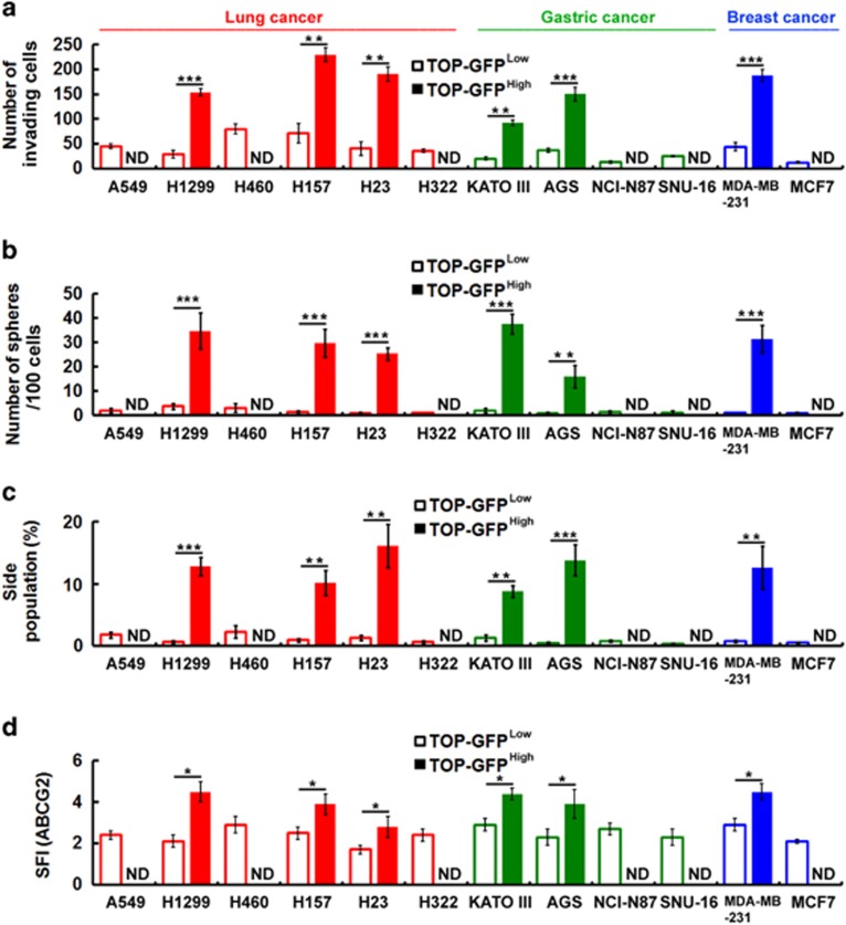
Figure 2.
Wnt signaling can promote the CSC phenotype in various types of cancer cell lines. (a–d) The highest and lowest 10% of TOP-GFP-expressing cell fractions were isolated by flow cytometry of TOP-GFP-transduced cell lines after treatment with Wnt3a-containing medium. Transwell invasion assays were performed to assess the invasive activity (a). The number of spheres formed was quantified (b). Cells were stained with Hoechst 33342. SP cells were counted (c). Cell surface expression of ATP-binding cassette (ABC)G2 was assessed by flow cytometry using anti-ABCG2 antibody (d). The specific fluorescence index (SFI) was calculated as the ratio of the geometric mean fluorescence value obtained with the specific antibody and the isotype control antibody. ND, not detectable. Data were derived from three independent experiments and are presented as the mean±s.d. *P<0.05; **P<0.01; ***P<0.005 (t-test).

