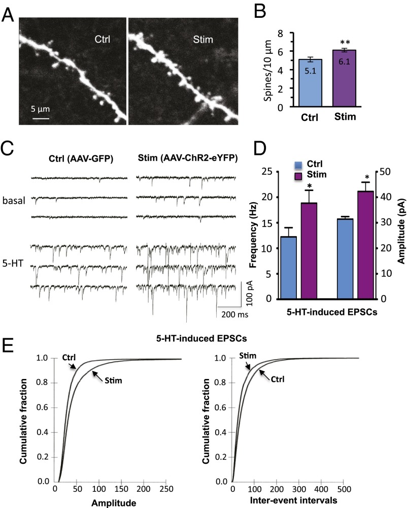
Fig. 5.
Optogenetic stimulation in vivo increases the number and function of spine synapses in IL-PFC pyramidal neurons. (A) Two-photon confocal z-stack projections of apical tuft dendritic segments of layer V pyramidal cells taken from rAAV2-GFP control virus or rAAV2-ChR2-eYFP–infused animals, both receiving in vivo light stimulation. (Scale bar, 10 μm.) (B) Spine density analysis; the results are the mean ± SEM (45 images from 11 cells from 5 rats for control; 66 images from 15 cells from 5 rats for stimulated; **P < 0.01; Student t test). (C) Examples of layer V pyramidal cell recording traces taken from control or stimulated animals; note the marked 5-HT–induced EPSCs in the cell taken from the stimulated animal. (D) Summary data showing both increased frequency (12.2 ± 1.8 and 18.8 ± 2.5 Hz, control and stimulated, respectively; P < 0.05) and amplitude (31.4 ± 1 and 42.3 ± 3.5 pA, respectively; P < 0.05) of 5-HT–induced EPSCs (*P < 0.05; Student t test; n = 15 neurons). (E) Cumulative probability distributions showing significant increases in amplitude (Kolmogorov–Smirnov two-sample test; P < 0.0000; z value = 12.9) and frequency (Kolmogorov–Smirnov two-sample test; P < 0.0000; z value = 10.9) of 5-HT–induced EPSCs (n = 15 neurons per group).

