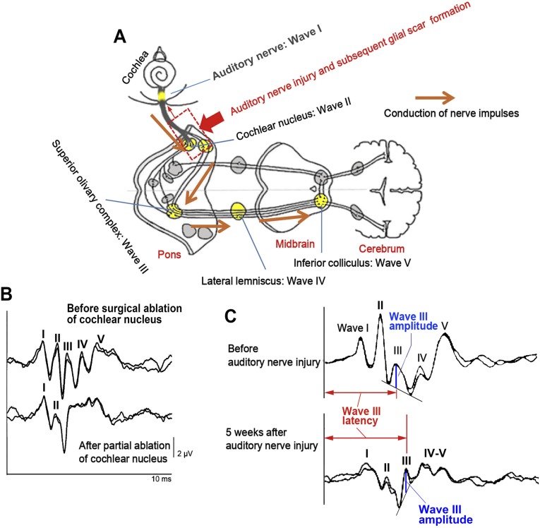
Fig. S2.
Neural generators of ABR and ABR waveform changes after surgical ablation of cochlear nucleus tissue and auditory nerve injury/subsequent glial scar formation. (A) In small experimental animals including rats, wave I of ABR is generated from the auditory nerve, and waves II, III, IV, and V mainly reflect synaptic activities in the cochlear nucleus, superior olivary complex, lateral lemniscus, and inferior colliculus, respectively. (B) Acute experiment to remove a part of the cochlear nucleus with a sucker depressed wave II and the following waves markedly while wave I maintained, confirming wave II was generated from the cochlear nucleus. Note the amplitude of wave I was not attenuated at this moment. (C) Normal configuration of ABR waveform was lost by auditory nerve mechanical injury and subsequent glial scar formation (dotted box in A). After compression, wave III was observed more apparently because of attenuation of wave II, the most prominent peak in small animal ABR. Measurements of wave III amplitudes and latencies are shown by blue perpendicular lines and red horizontal bidirectional arrows, respectively. Attenuation of wave I amplitude 5 wk after auditory nerve injury was probably due to retrograde and Wallerian degenerations of auditory neurons. Anatomical figure is cited from https://embryology.med.unsw.edu.au/embryology/index.php?title=File:Auditory_neural_pathway.jpg.

