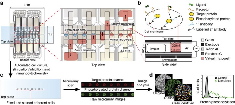
Figure 1. Digital microfluidic Immunocytochemistry in Single Cells (DISC).
(a) Top-view schematic of a digital microfluidic device used for cell culture, stimulation and immunocytochemistry. The expanded view illustrates the two primary fluid-handling operations, active and passive dispensing, required for metering and delivering reagents to cells cultured in virtual microwells. (b) Side-view schematic showing adherent cells cultured on the patterned top plate in a virtual microwell. The cells are sequentially treated with ligand and then probed for protein phosphorylation by immunocytochemistry. (c) Cartoon and microarray image illustrating how the top plate (a 3 by 1 inch slide bearing fixed and stained cells) is transferred from the device to a microarray scanner for laser scanning cytometry. (d) Images and data illustrating how the scans are processed by CellProfiler (open-source cell image analysis software) to identify individual cells and extract quantitative data. For each cell, the fluorescent intensity from the phosphorylated protein is normalized to the fluorescent intensity from the corresponding whole protein (phosphorylated+non-phosphorylated) to account for differences in expression level.

