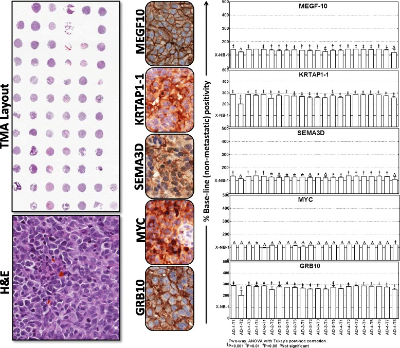
Fig. 5.

Localization and expressional modulation of metastamiRs target proteins in metastatic neuroblastoma. Representative image of the tissue micro array constructed with the replicates of non-metastatic xenografts controls and the manifold of metastatic tumors from spontaneous aggressive disease as well as the reproduced aggressive disease animals. Automated IHC stained panels showing the staining pattern and cellular localization of the metastamiRs’ protein targets (GRB10, MYC, SEMA3D, KRTAP1-1, MEGF10) in tumor samples. Histograms of Aperio-Spectrum image analysis and quantification of positivity for each target protein analyzed across the metastatic tumors of various animals presented with aggressive disease. The positivity values are compared to the non-metastatic xenograft controls using ANOVA with Tukey’s post-hoc correction using GraphPad PRISM
