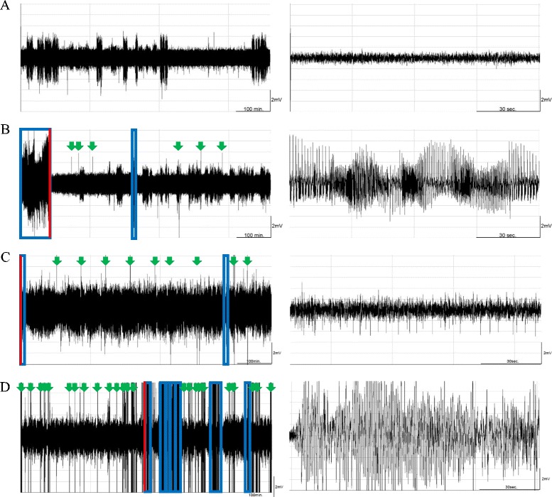
Fig. 1.

The effect of 10 Hz EA stimulation of bilateral Feng-Chi acupoints and naloxone on epileptic activities. Panels a, b, c and d respectively depict the EEG signals recorded from the naïve rats, the pilocarpine group, the PFS + EA + pilocarpine group and the naloxone + EA + pilocarpine group, beginning from the dark onset of the dark period. Pilocarpine was administered at time 0 in the left panels of b, c and d. The blue boxes represent the epileptiform EEGs. Red lines indicate the extracted time points for the expanded time-scale figures in the right panels. Green arrowheads are the artifacts. The larger amplitudes, with EEG signals less than 2 mV that appeared in panels a, were delta waves, which represent the state of slow wave sleep
