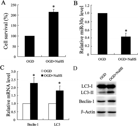
Fig. 3.

Effect of H2S on OGD-induced ischemic injury in SY-SH-5Y cell line. Cell model of OGD injury was established in hippocampal neurons-SY-SH-5Y cell line, which was injected with or without NaHS (100 μmol/l). (a) Cell viability was analyzed using MTT assay. (b) Quantitative analysis of expression of miR-30 profiles, (c) Beclin-1 mRNA and LC3 mRNA were examined by using Quantitative Real-Time PCR. (d) Expression of Beclin-1 and LC3-I/II protein were determined by using western blot assay. All values are expressed as the mean ± SD. * P < 0.05 vs. OGD group
