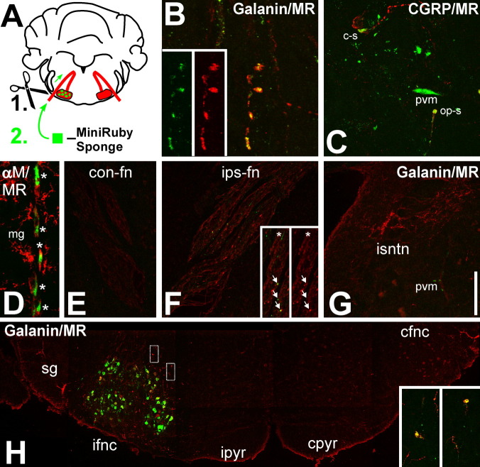
Figure 2.
Demonstration of central axonal sprout origin using Mini-Ruby, a dual anterograde/retrograde tracer. A: Schematic summary. A gelfoam sponge soaked in 1% Mini-Ruby solution was applied onto the fresh, proximal cut end of the facial nerve, followed by retrograde transport to axotomized motoneurons and a 14-day survival. B,C: High magnification of Mini-Ruby (green) colocalization with the immunoreactivity (IR) for the neuropeptides galanin (B) and CGRP (C) (red) in axonal sprouts just outside the axotomized facial motor nucleus. The double-labeled sprout in B and the bottom sprout in C are outward pointing (op-s); the top sprout in C is oriented in parallel to the center of the nucleus (“cruising,” c-s). The insets in B show the individual red and green fluorescence channels. In C, Mini-Ruby is also incorporated by perivascular macrophages (pvm). D: Mini-Ruby uptake in a string of alphaMbeta2 integrin (aM)-positive (red) perivascular macrophages (*) lining a cerebral blood vessel. The neighboring aM-positive and ramified microglia (mg) are devoid of Mini-Ruby. E–G: Double fluorescence for Mini-Ruby and galanin-IR in the descending intracerebral part of the contralateral facial nerve (E); the axotomized, ipsilateral facial nerve (F); and the ipsilateral spinal nucleus of trigeminal nerve (G, isntn). The insets in F show a higher magnification of the facial nerve (left, red and green; right, red only fluorescence). Note the double-labeled sprouts in the axotomized nerve (arrows) and their absence in the neighboring trigeminal nerve nucleus and contralateral nerve. The asterisk points to a Mini-Ruby+ but galanin– sprout. As in C, Mini-Ruby is frequently present in the populations of perivascular macrophages associated with larger blood vessels (D, pvm). The micrographs in E and F show the same galanin labeling motif as in Figure 1E,F but combine it with the Mini-Ruby fluorescence. H: Composite of Mini-Ruby and galanin-IR fluorescence in the ventral brainstem across the ipsilateral substantia gelatinosa, the ipsilateral and contralateral facial motor nuclei (ifnc, left; cfnc, right, respectively), and the ipsilateral and contralateral pyramidal tracts (ipyr and cpyr). Mini-Ruby neuronal cell body labeling is strictly limited to the axotomized facial motor nucleus, with a high density of galanin-positive sprouts in the surrounding tissue. Note the absence of both in the pyramidal tracts and the contralateral facial nucleus. The insets show higher magnifications for galanin/Mini-Ruby double labeling (yellow) of two sprouts just dorsal of the axotomized facial nucleus; their positions in the composite are indicated by the rectangles in H. A magenta/green version of Figure 2 is available as Supporting Information Figure 1. Scale bar = 10 μm in B, 45 μm in C and D, 270 μm in E–G; 350 μm in F insets.

