Figure 1.

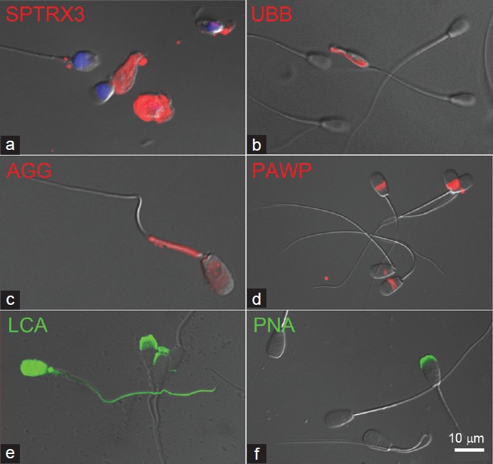
Fluorescent labeling of negative sperm quality biomarkers in human and animal spermatozoa. (a) Testis-specific thioredoxin SPTRX3 (red) is retained in the superfluous cytoplasm of defective human spermatozoa. (b) Ubiquitin (red) coats the surface of a defective bull spermatozoon with its flagellum coiled around the head but is undetectable in morphologically normal spermatozoa. (c) Aggresomes (red) are stress-induced aggregates of ubiquitinated proteins, here detected by ProteoStat kit in the mitochondrial sheath and head of a defective boar spermatozoon. (d) Sperm head postacrosomal protein PAWP (red) is detected at varied intensities in bull spermatozoa. (e) Lectin LCA (green) binds exclusively to the acrosomes of phenotypically normal bull spermatozoa but to the whole head and tail surface in the defective ones (segmental aplasia of the mitochondrial sheath is shown). (f) Lectin PNA binds to damaged/ruffled acrosomes, but not the intact ones in live bull spermatozoa. DNA in panel (a) was counterstained blue with DAPI. Epifluorescence micrographs are superimposed over parfocal images taken with DIC optics. PAWP: postacrosomal Sheath WWI Domain Binding Protein; LCA: Lens culinaris agglutinin; PNA: peanut agglutinin; DIC: differential interference contrast.
