
Fig. 1.
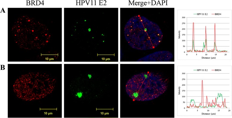
HPV replication foci in U2OS cells. Immunofluorescence analysis of U2OS cells transfected with HPV11 genome. U2OS cells were transfected with wt HPV11 minicircle genomes and grown on coverslips. Six days after transfection cells were fixed and immunostained with antibodies for HPV11 E2 (green) and BRD4 (red). Cell nuclei were detected by DAPI. Analysis was carried out at least three times starting from cell transfections. a and b represent cells with different number and size of HPV11 E2 foci. On the right panels, the intensity profiles are shown