
Fig. 18.
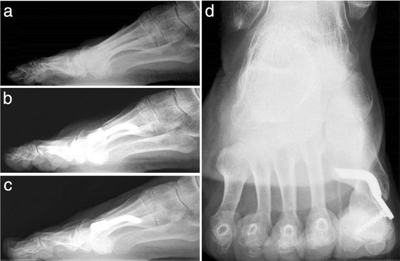
Case 12. A 71-year-old woman with moderate HV. a Preoperative lateral radiographic image. b At 1-month follow-up, lateral X-ray image. c Radiographic aspect at 48-month follow-up showing the maintained correction of the deformity and complete healing of the osteotomy. d X-ray image, sesamoid axial view, at 48-month follow-up